AI-Powered Medical Imaging Annotation Tool in Your Browser
Transform DICOMs into patient-specific 3D models, Collaborate in Real-time, and Train your own no-code AI Models-All Directly from Your Browser.
- Automatically generates patient-specific 3D visualizations
- Simplifies annotation of DICOMs
- Seamlessly fuses medical images
- Enhances diagnostic workflows
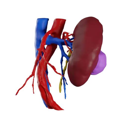
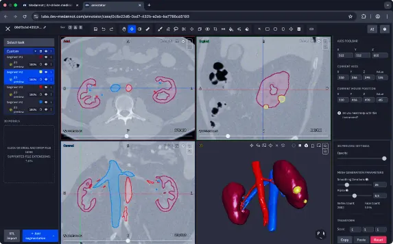
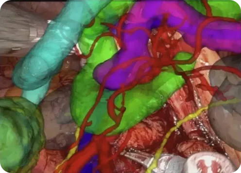
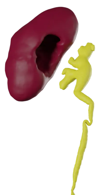
Patient-specific 3D kidney models—including tumors and vasculature. An essential tool in our preoperative planning.

Patient-specific 3D visualizations of facial bones from CBCT scans in minutes, used directly for intraoperative navigation.

Medannot allows us to go from a standard CT scan to a full 3D kidney model—including vessels and tumors—in under a minute.
Patient-specific 3D models from DICOM help surgeons plan better and operate with greater precision.
Patient-specific 3D kidney models—including tumors and vasculature. An essential tool in our preoperative planning.
Patient-specific 3D visualizations of facial bones from CBCT scans in minutes, used directly for intraoperative navigation.
Medannot allows us to go from a standard CT scan to a full 3D kidney model—including vessels and tumors—in under a minute.
Patient-specific 3D models from DICOM help surgeons plan better and operate with greater precision.
AI-Powered Annotation for Medical Imaging
Ready Wherever
You Are
Fully web-based, seamlessly integrates with hospital systems.
Built-In AI Model
Training
The AI model learns as you segment and label—no coding needed.
DICOM to 3D
Conversion
Generate patient-specific 3D models from DICOMs in seconds.
Collaborative
by Design
Seamless collaboration across clinical workflows in real time.
Built for Scale
& Research
Scalable platform with modular tools, APIs, and structured outputs.
Pre-Operative
3D Planning
Faster, seamless surgical planning with accurate, patient-specific models.
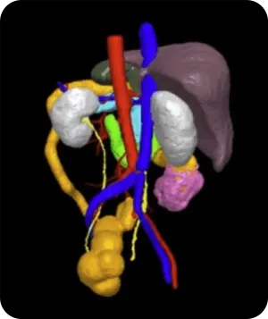
Transforming Medical Imaging into Actionable 3D Insights
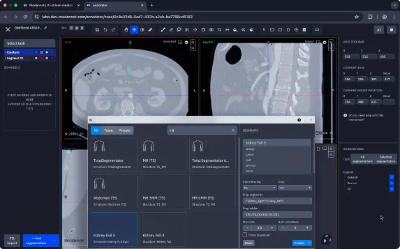
Convert DICOM to 3D Patient Models
Optimize Surgical Planning
Train Your Own AI — No Code Required
Use Medannot’s intuitive, web-based platform to build segmentation models without coding. Trusted by hospitals and researchers for generating fast, high-quality 3D datasets.
4 simple steps:
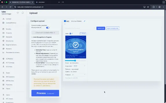
- Upload anonymized CT or MRI scans (DICOM format)
- Annotate using built-in or AI-assisted tools
- Train and deploy your custom model in a few clicks
- Generate tailored 3D visualizations for each patient
Accelerate Radiologic Diagnosis
Radiomics & Data Extraction
How Will Medannot Make My Job Easier?
Surgical Teams
& Surgeons
- Instantly create accurate 3D anatomical models for precise surgical planning
- Enhance intraoperative accuracy with detailed patient-specific insights
- Facilitate multidisciplinary team collaboration in real-time
Medical
Engineers
- Rapidly prototype and validate medical devices using accurate 3D models
- Collaborate seamlessly with clinicians to refine medical technologies
- Integrate precise anatomical data into engineering workflows
Radiologists
& Diagnostic Specialists
- Swiftly annotate and segment medical images with AI assistance
- Improve diagnostic precision and reduce interpretation time
- Generate patient-specific 3D visualizations effortlessly
AI & Machine Learning
Professionals
- Seamlessly annotate and label high-quality datasets
- Quickly train and deploy custom AI models without coding
- Validate segmentation models and iterate rapidly within a single platform.
Hospitals & Clinical
Research Institutions
- Centralize imaging workflows securely in the cloud
- Support compliance (FDA, MDR) and ensure data privacy
- Foster cross-departmental collaboration for diagnostics, research, and surgical planning
Medical Educators
& Academic Institutions
- Leverage interactive 3D modeling for effective teaching
- Provide realistic, hands-on learning experiences using actual patient scans
- Enhance educational outcomes in anatomy, radiology, and surgery
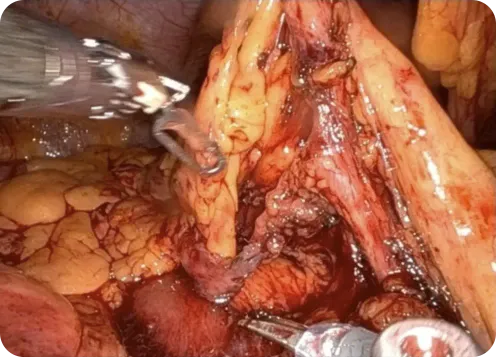
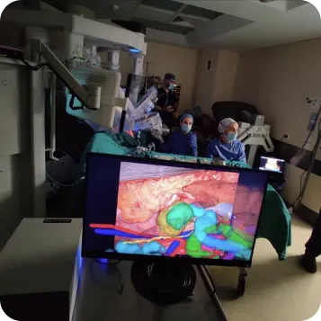
Case Study
Urology AI Solution at Ghent University Hospital
Segmentation Struggles in High-Stakes Surgical Planning
Urology surgeons at Ghent University Hospital faced significant challenges with manual segmentation of kidney structures, including tumors, arteries, and vessels. The manual process was time-consuming, prone to inaccuracies, and impractical for routine clinical use, hindering effective surgical planning.
Deployment of AI-Powered 3D Modeling
The surgical team utilized Medannot’s intuitive no-code AI platform to rapidly build and deploy their own custom AI segmentation model. The AI model precisely identifies and segments kidneys, tumors, and vascular structures directly from CT scans without the need for any programming or specialized IT skills.
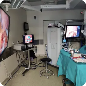
Turning Hours Into Minutes Where It Counts
The Medannot platform enabled Ghent’s surgical team to completely transform their workflow. What once required extensive manual labor and technical expertise can now be done in moments—directly by clinicians. This translated into measurable clinical impact:
- Immediate creation of accurate, patient-specific 3D kidney models
- Substantially improved precision in preoperative planning and surgical outcomes
- Complete elimination of dependence on coding, specialized hardware, and external technical support
- Streamlined workflows enabling surgeons to rapidly integrate 3D modeling into everyday practice
What Clinicians Say
Medannot allows us to go from a standard CT scan to a full 3D kidney model—including vessels and tumors—in under a minute. No engineers needed. No complicated software. Just clinical logic.
With Medannot, I created patient-specific 3D visualizations of facial bones from CBCT scans in minutes—used directly for intraoperative navigation. AI-powered and intuitive, it streamlined surgical planning like never before.
Medannot enables us to generate accurate, patient-specific 3D kidney models—including tumors and vasculature—with remarkable speed. It’s become an essential tool in our preoperative planning.
At Medannot, we empower our clinical colleagues with clearer insights into medical imaging. In modern practice, having comprehensive—and visual—understanding of both normal anatomy and pathology is essential. That’s why we built Medannot: to deliver fast, patient-specific 3D visualizations directly from DICOM data.

Enterprise-Ready Medical Imaging Annotation Tool


Collaboration Across Health Systems
Real-time, role-based collaboration across clinical teams and institutions.
Built for Healthcare-Grade Privacy
Encrypted and cloud-native. DICOMs are anonymized, with patient metadata stripped at upload.
Certified for Compliance
MDR & FDA ready, ISO and SOC 2 certified, and fully GDPR compliant.
Enterprise-Grade Infrastructure
Multi-team access, audit trails, and integrations for clinical and research deployment.
Experience Medannot
with a Free Trial
Discover how fast and intuitive AI-powered 3D modeling can be. Simply upload anonymized DICOMs, segment and label with your own AI, and generate patient-specific models in moments—right in your browser, no setup required.